Category:Animal embryos
Jump to navigation
Jump to search
earliest multicellular stage of development of an animal | |||||
| Upload media | |||||
| Instance of |
| ||||
|---|---|---|---|---|---|
| Subclass of | |||||
| Different from | |||||
| |||||
Subcategories
This category has the following 10 subcategories, out of 10 total.
Media in category "Animal embryos"
The following 149 files are in this category, out of 149 total.
-
 A lateral view 4 days post fertilisation zebrafish brain.jpg 4,263 × 2,387; 1.14 MB
-
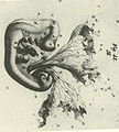
 A monograph on the development of elasmobranch fishes (1878) (14785187463).jpg 2,252 × 3,672; 427 KB
-

-
A-High-Throughput-Screen-for-Tuberculosis-Progression-pone.0016779.s004.ogv 1 min 12 s, 1,024 × 394; 3.55 MB
-
-
-
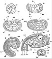
 Abatus cordatus Developmental stages.jpg 1,311 × 1,679; 1.1 MB
-
 Abatus cordatus Hypothetical representation of perigastrulation.jpg 682 × 711; 449 KB
-
 Abatus cordatus Long-spined juvenile (J2) (01).jpg 1,032 × 1,402; 1.43 MB
-
 Abatus cordatus Long-spined juvenile (J2).jpg 879 × 1,080; 781 KB
-
 Abatus cordatus Post-gastrular stage 100 days after fertilization.jpg 1,025 × 1,566; 1.41 MB
-
 Abatus cordatus Post-gastrular stage.jpg 930 × 1,284; 1.04 MB
-
Accessing-the-Microscopic-World-pbio.0030012.sv001.ogv 1 min 49 s, 240 × 180; 7.14 MB
-
 Amphioxus Growth of the ciliated embryo.jpg 1,735 × 2,138; 1.26 MB
-

-
-
-
-
-

-
 Aves Clustering of seemingly stochastic EMT underpins the formation of PS.jpg 780 × 1,500; 174 KB
-

-
 Aves Development from 1 tot 41 somites.jpg 1,646 × 1,768; 1.65 MB
-

-
 Aves Diagrams depicting the early stages of chick development.jpg 1,500 × 922; 152 KB
-
-
-
 Aves Embryo of aboul 27 somites drawn in alcohol by reflected light; upper side, x 10.jpg 1,287 × 1,385; 1.68 MB
-
 Aves EMT in the formation of the primitive streak.jpg 856 × 1,500; 351 KB
-
 Aves Entire embryo of 35 s.jpg 936 × 1,186; 1.69 MB
-
 Aves FGF signalling in mesoderm migration.jpg 2,152 × 612; 371 KB
-
 Aves gastrulation Mechanism of cell ingression.jpg 1,478 × 1,127; 175 KB
-
 Aves gastrulation Origins of flow organisation.jpg 1,660 × 1,579; 661 KB
-
 Aves gastrulation Tissue flows driving primitive streak formation.jpg 1,663 × 1,621; 710 KB
-

-
 Aves Neural tube and somites in chick embryo.jpg 1,472 × 1,144; 674 KB
-
 Aves Surface views of two stages of the blastoderm of the egg of the sparrow.jpg 1,456 × 789; 883 KB
-
 Aves Three stages of the blastoderm to show the extension of the mesoblast.jpg 1,486 × 626; 885 KB
-
 Aves Transverse section through the seventeenth somite of a 29 s embryo..jpg 1,210 × 730; 952 KB
-
 Aves Transverse section through the twentieth somite of a 29s embryo.jpg 1,920 × 859; 1.4 MB
-
 Aves Transverse section through the twenty-ninth somite of a 29 s embryo x10.jpg 1,161 × 1,358; 2.55 MB
-
 Aves Transverse section through the twenty-ninth somite of a 29 s embryo.jpg 1,375 × 741; 1.13 MB
-
 Aves Transverse section through the twenty-sixth somite of a 29 s embryo.jpg 1,363 × 731; 1.33 MB
-
 Avian development before gastrulation.jpg 2,767 × 3,354; 1.41 MB
-
 Bischoff Vorlage.jpg 1,616 × 1,789; 1.24 MB
-
 Cell flow patterns during streak formation for different mechanisms.jpg 1,929 × 1,046; 1.7 MB
-
 Cnr1 Expressionsmuster.jpg 501 × 790; 81 KB
-
CXCR7-Functions-as-a-Scavenger-for-CXCL12-and-CXCL11-pone.0009175.s001.ogv 6.3 s, 335 × 311; 243 KB
-
CXCR7-Functions-as-a-Scavenger-for-CXCL12-and-CXCL11-pone.0009175.s002.ogv 5.7 s, 321 × 340; 186 KB
-
 Diagrams showing the development of the amnion, chorion and allantois.jpg 1,269 × 1,151; 605 KB
-
-
-
-
-
-
-
-
-
-
-
-
-
-
-
-
-
Early-Endocardial-Morphogenesis-Requires-SclTal1-pgen.0030140.sv001.ogv 12 s, 720 × 480; 5.04 MB
-
Early-Endocardial-Morphogenesis-Requires-SclTal1-pgen.0030140.sv002.ogv 8.7 s, 720 × 480; 1.43 MB
-
Early-Endocardial-Morphogenesis-Requires-SclTal1-pgen.0030140.sv003.ogv 8.3 s, 720 × 480; 724 KB
-
 EB1911 Lamellibranchia - development of Ostrea edulis.jpg 775 × 985; 261 KB
-
 EB1911 Lamellibranchia - embryo of Pisidium - diagram.jpg 405 × 439; 90 KB
-

-
 EB1911 Lamellibranchia - embryo of Pisidium pusillum - surface view.jpg 378 × 477; 101 KB
-
 EB1911 Lamellibranchia - embryo of Yoldia limatula.jpg 969 × 400; 106 KB
-
 EB1911 Peripatus - P. capensis - Series of Embryos.jpg 952 × 311; 78 KB
-
 EB1911 Peripatus - Series of Embryos.jpg 1,011 × 723; 218 KB
-
 EB1911 Tunicata - Stages in the Embryology of a Simple Ascidian.jpg 805 × 913; 216 KB
-
 Embryonic development of the chondrocranium of a lizard.jpg 343 × 800; 140 KB
-
-
Fast-imaging-of-live-organisms-with-sculpted-light-sheets-srep09385-s1.ogv 4.1 s, 960 × 540; 4.04 MB
-
Fast-imaging-of-live-organisms-with-sculpted-light-sheets-srep09385-s2.ogv 4.1 s, 960 × 540; 2.61 MB
-
Fast-imaging-of-live-organisms-with-sculpted-light-sheets-srep09385-s3.ogv 4.1 s, 960 × 540; 2.93 MB
-

-
-
-
-
 Hand-book of physiology (1892) (14765113152).jpg 1,252 × 1,100; 313 KB
-
-
 Heilmann fig60.jpg 900 × 858; 259 KB
-
-
Imaging-multicellular-specimens-with-real-time-optimized-tiling-light-sheet-selective-plane-ncomms11088-s15.ogv 11 s, 1,440 × 1,080; 5.15 MB
-
Imaging-multicellular-specimens-with-real-time-optimized-tiling-light-sheet-selective-plane-ncomms11088-s16.ogv 8.0 s, 1,200 × 608; 2.55 MB
-
-
-
Imaging-multicellular-specimens-with-real-time-optimized-tiling-light-sheet-selective-plane-ncomms11088-s8.ogv 40 s, 1,248 × 960; 19.49 MB
-
-
-
-
-
-
-
-
 Louisiana Pearshell Embryos (5563959741).jpg 3,968 × 2,976; 2.68 MB
-
-
-
-
-
-
-
Multiplexed-3D-FRET-imaging-in-deep-tissue-of-live-embryos-srep13991-s2.ogv 36 s, 610 × 470; 1.14 MB
-
Multiplexed-3D-FRET-imaging-in-deep-tissue-of-live-embryos-srep13991-s3.ogv 35 s, 610 × 270; 512 KB
-
Multiplexed-3D-FRET-imaging-in-deep-tissue-of-live-embryos-srep13991-s4.ogv 36 s, 910 × 420; 1.58 MB
-
Multiplexed-3D-FRET-imaging-in-deep-tissue-of-live-embryos-srep13991-s5.ogv 24 s, 616 × 602; 1.03 MB
-
Multiplexed-3D-FRET-imaging-in-deep-tissue-of-live-embryos-srep13991-s6.ogv 24 s, 686 × 672; 1.41 MB
-
Multiplexed-3D-FRET-imaging-in-deep-tissue-of-live-embryos-srep13991-s7.ogv 24 s, 1,190 × 602; 2.05 MB
-
Multiplexed-3D-FRET-imaging-in-deep-tissue-of-live-embryos-srep13991-s8.ogv 24 s, 1,190 × 602; 2.13 MB
-
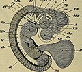
 Pig embryo median sagittal section.jpg 1,257 × 1,143; 617 KB
-
 Pig embryo transverse section.jpg 889 × 772; 397 KB
-
-
-
 PZSL1889Page129.png 1,752 × 2,854; 4.53 MB
-
 PZSL1889Plate46.png 1,903 × 3,026; 5.5 MB
-
 PZSL1889Plate49.png 1,913 × 2,979; 5.66 MB
-
 PZSL1889Plate59.png 1,845 × 2,942; 4.76 MB
-
-

-

-
 Section of squid embryo stained with stained with Toluidin Blue Light micrograph.jpg 1,410 × 3,000; 796 KB
-
 Section of squid embryo stained with Toluidin Blue.jpg 1,410 × 3,000; 810 KB
-
 Semisulcospira libertina 2.jpg 701 × 701; 157 KB
-
 Spiral cleavage in Trochus.png 2,067 × 629; 609 KB
-
Sumoylation-of-CCAATenhancer-binding-protein-α-is-implicated-in-hematopoietic-stemprogenitor-cell-srep09011-s2.ogv 14 s, 1,920 × 1,080; 2.68 MB
-
Sumoylation-of-CCAATenhancer-binding-protein-α-is-implicated-in-hematopoietic-stemprogenitor-cell-srep09011-s3.ogv 14 s, 1,920 × 1,080; 2.58 MB
-
 The development of the chick; an introduction to embryology (1908) (14725950606).jpg 1,494 × 2,220; 478 KB
-
-
-
-
-
-
-
-
-
-
-
-

-
Wnt5-signaling-in-vertebrate-pancreas-development-1741-7007-3-23-S1.ogv 22 s, 320 × 240; 133 KB
-
Wnt5-signaling-in-vertebrate-pancreas-development-1741-7007-3-23-S3.ogv 18 s, 320 × 240; 145 KB
-
 Развитие эмбриона аквариумной рыбки.jpg 2,048 × 783; 112 KB

_(14785187463).jpg)


_(01).jpg)
.jpg)


_(14764634251).jpg)


_of_primitve_streak.jpg)


.jpg)
_(14765113152).jpg)

.jpg)


_and_median_sagittal_sections_(g_and_h)_using_the_rabbit_as_a_model.jpg)


_(14725950606).jpg)
_(14756798946).jpg)